- Suite 1335, 13/F.,
Central Building,
1-3 Pedder Street,
Central, Hong Kong MTR
Central Station (Exit G) - 3104 0802


- |


- |

-
Suite 1335, 13/F.,
Central Building,
1-3 Pedder Street,
Central, Hong Kong MTR
Central Station (Exit G) - 3104 0802
- Home
- Venous Disease

Venous Disease
靜脈曲張
Venous Disease
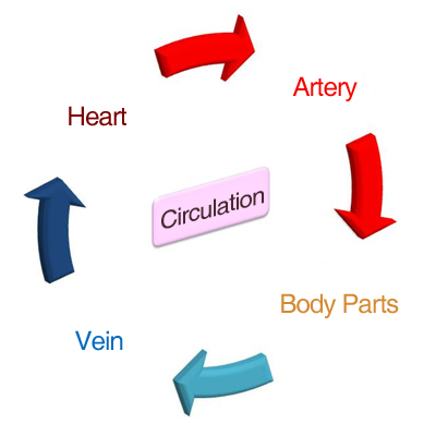
Heart pumps oxygen-rich blood through a network of arteries to provide the needed nutrient and oxygen to all body parts. Veins return blood from body tissue to be enriched with oxygen again and re-pumped by the heart. Essentially, blood flows down our legs through arteries, and up to the legs via veins back to the heart.
 |
One-way flow
|

- Open passage
- One way valve
- Muscle pump
Cause of chronic venous disease
There are two systems of veins: deep veins which are embedded inside muscles and superficial veins which are just underneath our skin. Blood drains from superficial veins through perforating veins into deep veins which then provides pathways to the heart.
During walking as muscle contract blood inside the deep vein is squeezed and pushed upward. Valves inside the veins open as blood flows towards the heart and close to stop blood from flowing back towards the feet.
When wall of the veins are weak, the leaflets of the valves can no longer meet properly to stop back flow of blood causing congestion. As a result, blood pools and superficial veins puff up as varicose veins or spider veins. The increased pressure inside the veins may cause swelling and dragging pain to our legs. The abnormally high pressure also elicits a cascade of inflammatory events and leads to darkening of skin, eczema and even non-healing ulcer.
Obstruction of the deep veins is another less frequent cause of venous problems
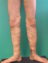
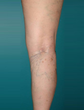
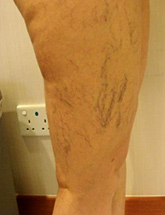
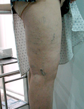
What are varicose veins?
If valves at the junction between the deep veins and the superficial veins are not closing properly, blood is allowed to flow backward from deep veins to superficial veins. Hence, superficial veins are distended and lengthen causing them to bulge out and become tortuous. Origin of the abnormal back-flow is usually at the groin with blood reflux along the great saphenous vein causing varicose veins in the inner aspect of the thigh and calf. Another common location of the incompetent valve is at the back of knee with back-flow along the small saphenous vein and varicose veins at the back of the calf. However, any of the perforating veins between the deep veins and the superficial veins can be incompetent and give rise to groups of varicose veins at other location along the lower limb
How are varicose veins diagnosed?
- Accurate identification of the origin of back flow (reflux) is a prerequisite for the planning of any intervention. Varicose vein is diagnosed primarily by physical examination. The usual pattern of distribution are the
Great Saphenous Vein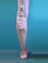
Small Saphenous Vein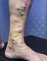
Perforating Vein
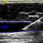
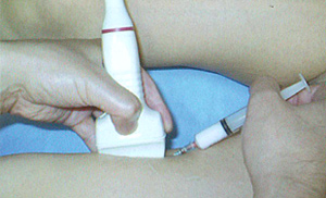
- Reflux often can be deduced by physical examination and confirmed by Duplex scan.
Duplex scan is:
- A special ultrasound scan with the additional ability to measure blood flow direction.
- Detects leaking valves, direction of blood flow and evidence of blood clots.
- The non-invasive test of choice to locate the source and channel of abnormal backflow of blood.
- Precisely pinpoint the origin of abnormal back-flow, provides a map of the affected veins and their connection.
- It is indispensable in tailoring treatment to each individual. Has to be performed before contemplating any intervention.
Is treatment of Varicose vein medically necessary?
- Stagnation of blood causes distending dragging pain, easy fatigue, cramp and leg swelling. The abnormally high pressure inside the superficial venous system also elicits a cascade of inflammatory events and leads to darkening of skin, eczema, bleeding and even chronic non-healing ulcer. These can be prevented when the incompetent veins are sealed off. Surgical interventions are medically indicated to relief symptom and prevent complication.
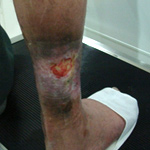
Non-healing Ulcer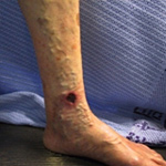
Non-healing Ulcer
Stasis Eczema
Stasis Eczema
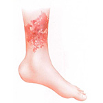
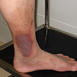
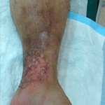
Non-healing Ulcer Stasis Eczema
What causes varicose vein?
Hereditary is the number one contributing factor. Hormonal factors include pregnancy. Other predisposing factors are standing occupations, obesity and heavy weight lifting.
What you can do?
You can't do anything about your heredity, age, or gender. However, you can help delay the development of varicose veins or keep them from progressing.
Conservative management of venous diseases
- Avoid over-weight
- Avoid obstructing the veins. Do not wear tight socks, girdles or garters
- Avoid high-heel shoes as it prevent calf muscle contraction
- Avoid prolonged standing or sitting still. Change posture, flex your feet or tip toes 2-3 times every few minutes to contract the calf muscle pump
- Avoid crossing legs while sitting
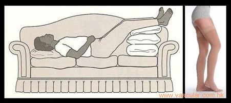
- Patient with severe symptom, benefit from leg elevation above the level of heart for 1/2 hour, three times daily. Leg elevation through-out the night for patient with significant edema
- Wear correctly fitted graduated compression elastic stocking. Replace when they become loose-fitting
- See a surgeon who can diagnose the cause of your varicose veins, the sources of venous reflux
What are the treatment options?
Apart from their non-appealing appearance, they may cause aching pain and swelling legs; and may result in serious complications such as skin discoloration, non-healing leg ulcers and blood clots. Early treatment alleviates symptom and prevents advance complications.
Highly effective current treatment options to close or remove abnormal veins include sclerotherapy, minimally invasive endovenous ablation, surgical vein stripping and phlebectomy.
The extent and distribution of the disease are crucial in choosing the treatment options. Feel free to discuss the benefits and side effects with a vascular surgeon.
- Stockings are effective in controlling symptoms but are only effective if worn regularly. In hot environments, the use of elastic stockings may be impractical.
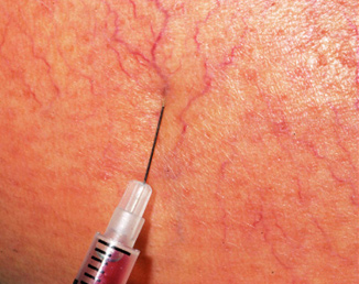
- Injection Sclerotherapy
- A friendly procedure which can be performed in a clinic. Medicine is injected into the spider veins or small varicose veins to close the abnormal veins and allows blood to re-route through normal veins
- Offers the best results in limbs with localized areas of reflux.
- Sclerosant in the form of microfoam is more effective for large varicosity.
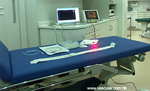
- Endovenous ablation (EVLT / RFA)
- Techniques that seal off the incompetent (abnormal) saphenous trunk (Great or Short) from the inside using either using chemical drugs or heat technology (Laser or radiofrequency).
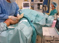
- These through the lumen of the vein procedures are performed under local anaesthesia.
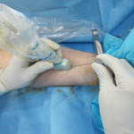
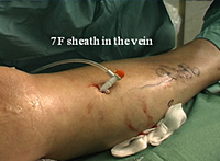
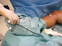
- With you lying down ultrasound scan will be used to locate the abnormal vein. The vein is punctured through the numbed skin to introduce a very thin tube (catheter) into the main trunk of the superficial venous system
- Through the catheter a fibre is inserted. Laser energy or radiofrequency energy is then delivered via the fibre.
- As the fibre is withdrawn the vein is shrunken and the inner tissue of the vein is destroyed. The healing process of the body then seals off the abnormal veins and re-routes blood flow to normal functioning veins.
- Shortly after the procedure you can walk as usual and go home. Resume daily activities in the same day.
- Endovenous Laser Therapy (EVLT)
- The skin is numbed, under ultrasound guidance, a catheter is inserted through a needle stick into the abnormal vein.
- Endovenous RFA
- Similar to EVLT but radiofrequency energy is used
- Similar to EVLT but radiofrequency energy is used
- Biochemical Glue (Venaseal)
- Under ultrasound guidance the abnormal vein is punctured
- A fine long tube is introduced into the main trunk of the superficial venous system.
- Small aliquots of bio-compatible glue are deposited along the vein to stick the wall closed.
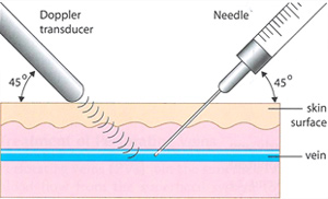
- Ultrasound guided sclerotherapy
- Abnormal superficial veins that are slightly deeper beneath the skin may not be visible.
- Ultrasound scan has to be used to visualize the abnormal vein.
- The vein is punctured under direct guidance of the ultrasound view.
- Sclerosing agent is then injected to seal off the vein
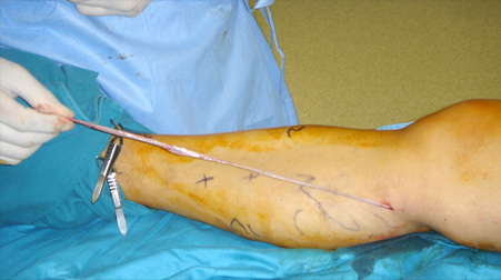
- Open Surgery is the traditional way of treating varicose vein and is performed under local or general anaesthesia. It involve several steps and patients are able to resume normal activities after a few hours
- Ligation: The origin of the abnormal backflow of blood is located by ultrasound. Under ultrasound image control, a small skin incision is made to tie off the origin at the junction between the superficial vein and the deep vein.
- Invagination stripping: The channel of reflux is removed (stripped). A "PIN stripper" is inserted through the cut end and advanced along the main trunk of the superficial vein. The end of the PIN stripperis sewn to the end of the vein. The tip of the stripper is perforated through the skin at knee level and the stripper is removed. The vein is pulled in on itself and is "stripped" stripped inside-out through a 2mm puncture wound at just below the knee level
- Hook phlebectomy: Through multiple fine stab incisions a hook is inserted to fish out and remove the tributary varicose veins. Scarring is minimal.

How to choose?
- Patients are attracted to the simplicity of the minimally invasive endovenous procedures with its post surgery's immediate mobilisation and short recuperation period.
- Though endovenous procedures are prevalent in treating patients with varicose veins, yet it still cannot replace open surgical procedures in all situations.
- Treatment methods are not mutually exclusive.
- Depending on the distribution and extent of the varicose veins, treatment methods are often combined to achieve optimal results with the least invasiveness.
- Endovenous ablation is used to treat the main trunk (i.e the Great Saphenous Vein, or the short saphenous vein). The fibre used for endovenous ablation is flexible but too rigid to be threaded along tortuous branches of varicose veins. After the main trunk is treated, the large branch varicose veins are hooked out and removed through puncture holes of 2mm.
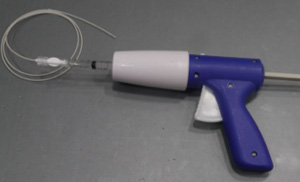
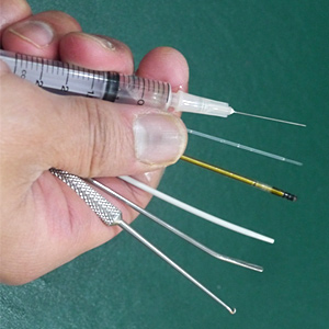
-

- Injection needle
- Laser fibre
- Radiofrequency fibre
- Glue catheter
- PIN Stripper
- Hook
- Injection needle
- Laser fibre
- Radiofrequency fibre
- Glue catheter
- PIN Stripper
- Hook
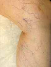
What are spider veins?
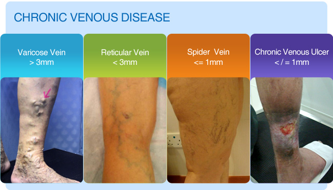
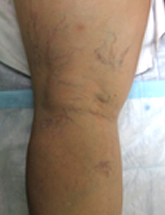
- Veins become congested with blood resulting in dilated abnormal veins. There are three types of abnormal veins, which are frequently seen in combination. "spider veins" (telangiectasias) are the fine red or purple capillary veins on the surface of the skin. The larger blue veins are called "reticular veins", and are slightly deeper below the skin's surface. Varicose veins are the largest of the abnormal superficial veins. Varicose veins are tortuous veins that are located somewhat deeper than the spider veins and bulge above the skin's surface.

What are the treatments for spider vein?
Backflow from larger veins into smaller capillaries causes their distension and the formation of spider veins. Underlying abnormal back flow of blood and varicose veins should be treated before treating the spider veins.
- Sclerotherapy is the treatment for the small reticular veins and spider veins. Using very fine needle the veins are injected with sclerosant that irritates the inner lining, causes mild inflammation, obliteration, and subsequent reabsorption of the unwanted veins. The treated veins will fade as the body gradually removes them.